A camera builds a surface model
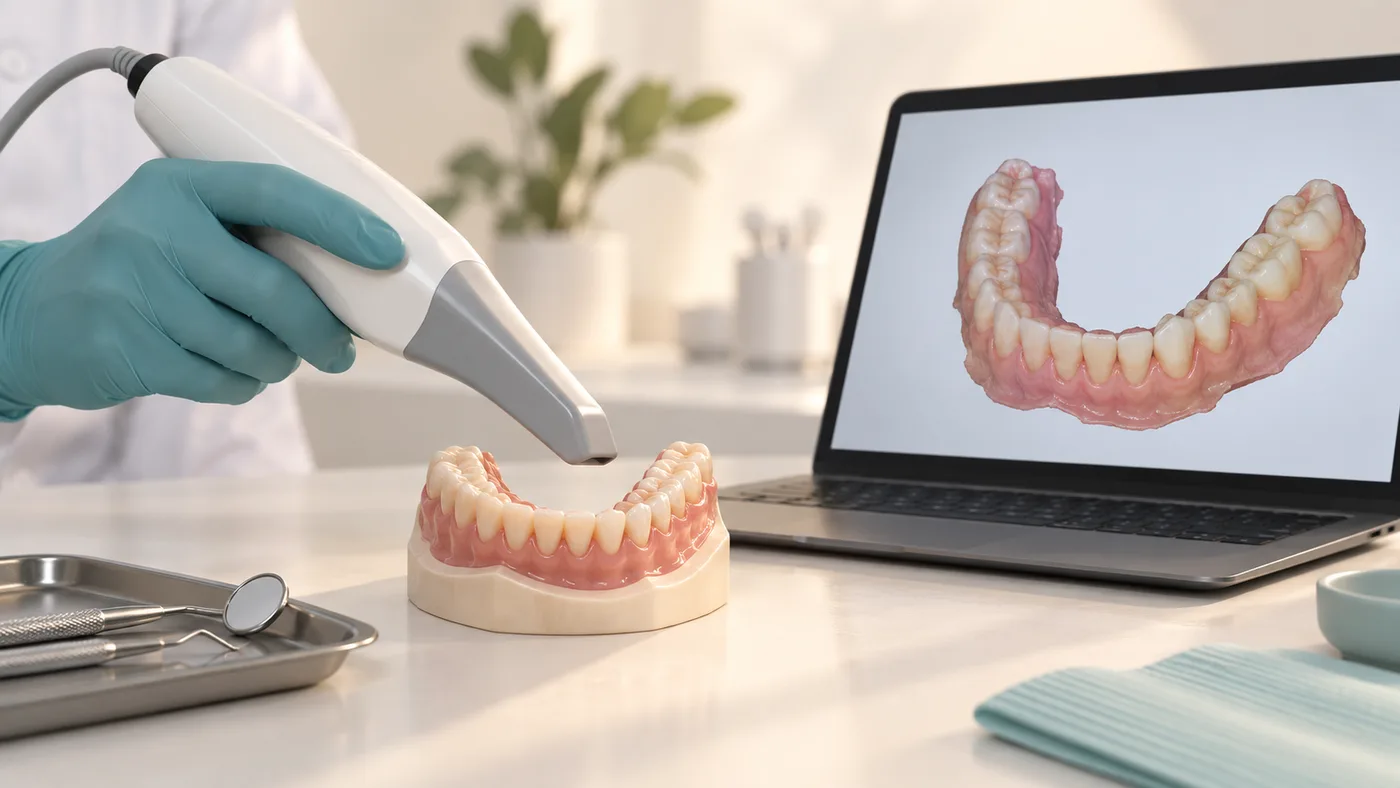
An intraoral scanner records visible tooth and soft-tissue surfaces and combines many views into a three-dimensional model. It does not see through a tooth, identify every cavity or show the jawbone like an X-ray examination.
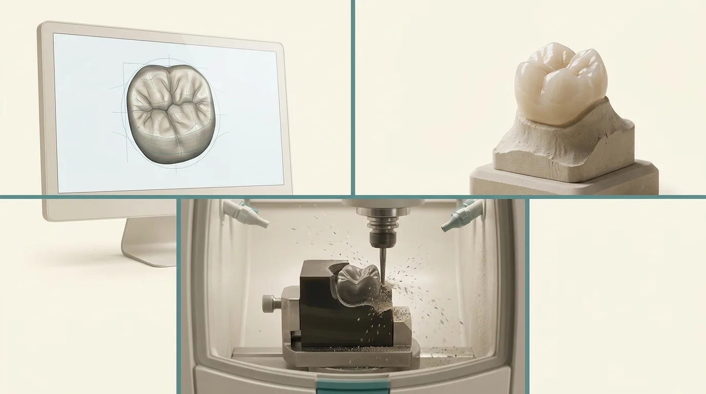
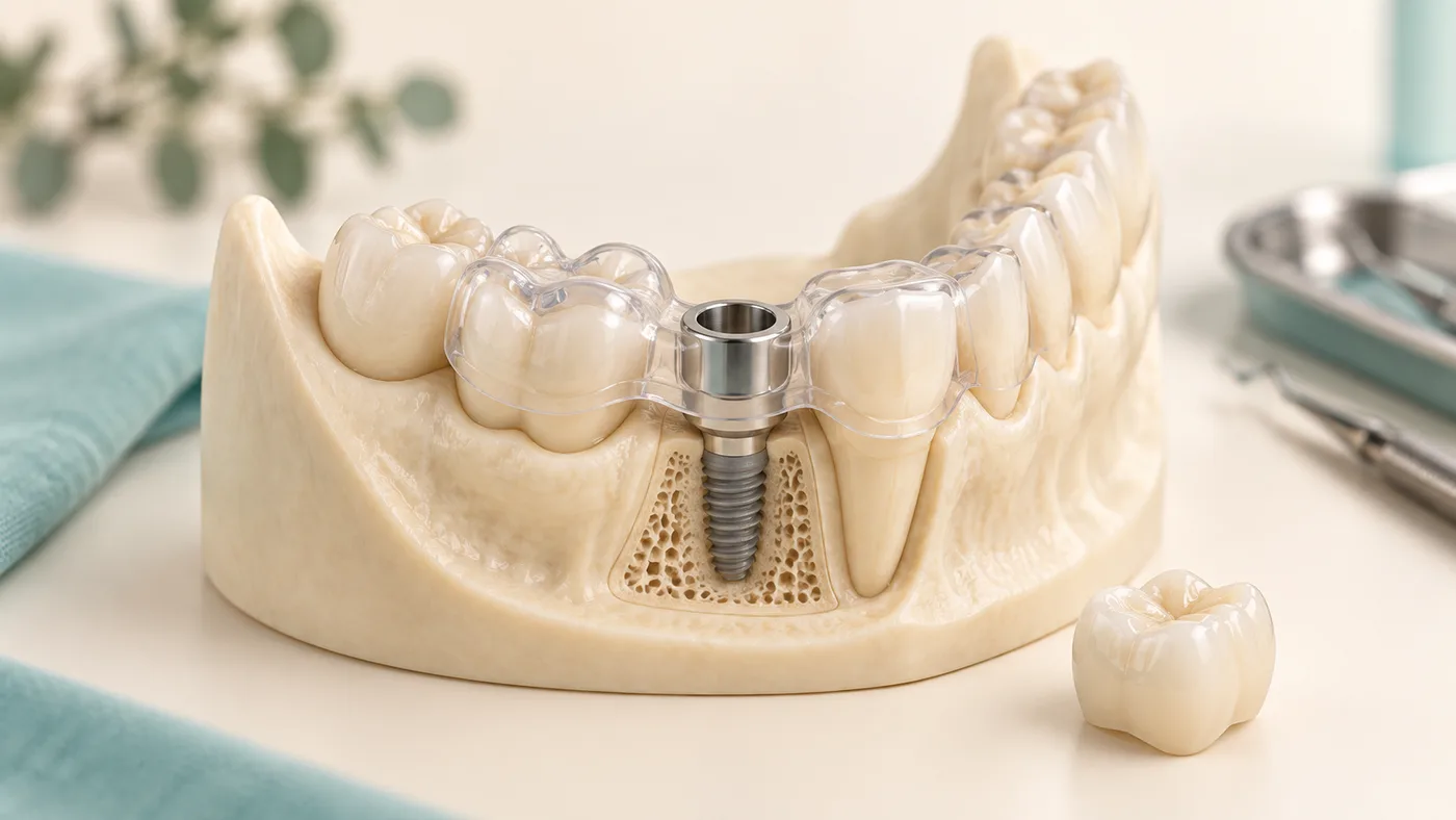
The model can support crown design, orthodontic planning or selected implant-restoration workflows. Accuracy depends on a suitable technique, moisture control, visible margins and the clinical situation. A scan that looks attractive on screen is not automatically complete.
What happens during the scan?
A small scanning head moves around the teeth. The team may gently retract the cheek, dry a surface and record the opposing arch and bite. Missing areas can sometimes be captured again locally. Patients who dislike impression material may find this easier, although a scanner can still be uncomfortable with limited opening or a strong gag reflex.
Where conventional records remain useful
Moving tissues, deep margins, blood or saliva can obscure the area needed for a restoration. Recording a completely toothless arch also involves tissue support and function that a simple surface scan may not capture adequately. Some extensive implant cases require additional verification. A conventional or combined method can therefore be the better clinical choice.
Why the bite record matters
A precise-looking tooth shape is only part of the task. The laboratory also needs the relationship between the jaws and clear instructions about the planned restoration. Errors in this information can affect contact points, bite and comfort even when the scan itself is detailed.
Can an old scan be reused?
A stored scan can provide a useful comparison. It cannot automatically replace new records after teeth move, restorations change or tissues heal. Ask what the scan will be used for, whether additional records are needed and how the team will check the finished restoration in your mouth.