
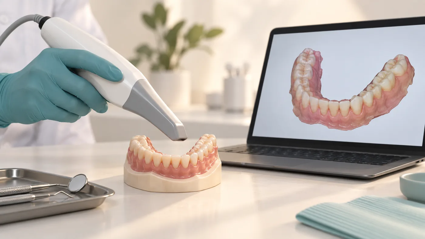
A digital record of visible surfaces
An intraoral scanner captures the shape of teeth and accessible soft tissues. The resulting surface model can help plan or manufacture selected crowns, aligners, and other restorations.
Where it helps—and where it needs checking
A scan can be reviewed immediately and missing areas recaptured. However, saliva, bleeding, movement, deep margins, reflective surfaces, and long scan spans can affect the record. Completely toothless arches and mobile tissues may need conventional impressions or a combined workflow.
It is not an X-ray
An optical scan does not show hidden roots, internal decay, or jawbone in the way a radiographic examination can. Your clinician selects additional records when the diagnosis requires them.
For extensive implant work
The team must verify that the captured implant positions and bite are sufficiently reliable for the planned restoration. A detailed-looking model alone does not demonstrate a passive, accurate fit.